Live and in Color: Why Color Calibration is So Important in Medical Technology
In the areas of microscopy, dermatology and ophthalmology, digitally captured images are particularly effective aids in the diagnostic process. The color is often an important criterion to evaluate whether a structure is healthy or pathological.

Color calibration for Basler cameras
In our white paper, we explain why the calibration of Basler color cameras is important and outline the key steps involved
What is meant by color and what color systems are there?
What are the advantages of the RGB color space?
What benefits does a calibrated camera offer?
What are the four steps of color calibration at Basler?
In our article “What Role Does Color Play in Image Processing?” we already explained the role of color reproduction in machine vision. While monochrome images often suffice for industrial image processing, very different types of image information are used in Medical and Life Sciences, which is why color fidelity plays such an important role here.

Color in ophthalmology
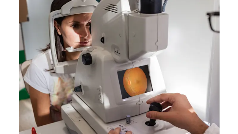
Let’s take a closer look at ophthalmology. Digital fundus cameras are used to examine the retina. The physician positions the camera in front of the patient’s eye and captures an image. This high-resolution image is then examined with the help of analytical software. Based on these images, the physician can detect whether the thin vessels in the retina are fully connected and intact or, if that’s not the case, whether additional exams are required. This type of diagnostic process helps prevent diseases such as macular degeneration.
Highly reliable color rendering as well as the reproducibility of the images is particularly important here since the color provides information about the condition of certain tissues. For example, tissue can discolor when it doesn’t receive an adequate supply of oxygen.
The camera must be calibrated properly to ensure the reliability of the color rendering.
Color calibration of cameras
Optimizing the parameters within the color computation pipeline in the camera’s firmware is referred to as the color calibration. A so-called color defect is the basis of the calibration. A ColorChecker is normally used in the calibration process. The ColorChecker is a chessboard-style card with 18 colors and 6 gray shades depicted as squares next to one another. When viewed under certain defined lighting, the sRGB values for the individual fields are a known factor, allowing the ColorChecker to serve as a reference benchmark for the colors as detected by the camera.
The color value for the camera is measured based on a specific field, providing the sRGB value that can then be used to compare with the actual, known value for the color field. The difference between the measured and known point in the color space is noted as the so-called color defect ΔE. The goal of the calibration process is to parameterize the individual function blocks in the camera’s color pipeline in such a way that the color defect ΔE is minimized compared to the ColorChecker’s reference values.
After the color calibration, it is possible to perform additional modifications of individual colors ‘against’ the color defect to potentially take existing color profiles into account. This is particularly true for applications in which consumer cameras are replaced by industrial cameras.
Industrial cameras offer great advantages in color depiction compared to cameras in the consumer field. This is due to the fact that the color pipeline in consumer cameras is a black box that usually can’t be parameterized. In those cases, the color pipeline is designed to generate aesthetic images – and not to depict reality as precisely as possible. For that reason, industrial cameras with enhanced color features are preferred for applications specifically in medical imaging and diagnostics.
Trends
The color depiction of cameras is continuously improving. Trends show that applications in which traditionally only monochrome sensors were used are now also operated with color cameras (e.g., in fluorescence microscopy). In other words, cameras are becoming more multifunctional. Cameras with special color features for Medical and Life Sciences offer developers function blocks (color pipeline) that can be parameterized in the camera’s firmware.
Our products for image processing in medicine
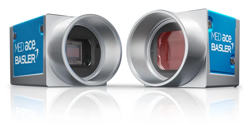
Benefit from the specially designed Basler MED ace camera series along with our experience in developing individual machine vision solutions for your requirements.
