Examine Cell Processes in Microscopic Detail
ace camera: a sharp and quick eye for cell research
- Customer
- SYNENTEC GmbH
- Location
- Elmshorn, Germany
- Date
- 2013
Research is important when it is about deciphering the mechanism of a disease. SYNENTEC GmbH develops and produces imaging microscopes for fully automatic cell analysis. NYONE is a device that is used primarily in cell research and drug discovery. Using NYONE, it is possible to obtain a detailed view of the cells' interior, resolving even the smallest structures.
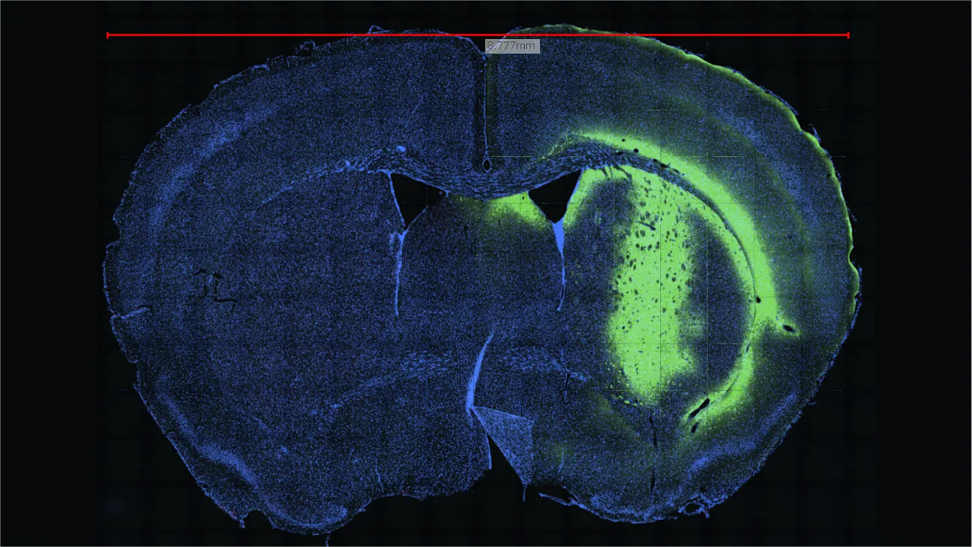
Image processing provides the details for microscopic analysis
NYONE helps us understand processes within the cell, maps its tracks digitally, and converts them into comprehensible data. Image processing and digital cameras are irreplaceable tools in this endeavor.
Metabolic processes and defects in cells
Image-based cytometry using NYONE allows us to have a deep insight into the processes of life. With resolutions from millimeters to the nanometer scale, NYONE gives us a better understanding of the metabolic processes in cells.
The challenge was to find a camera that delivers high-quality images at a high data rate. This is a must for microscopic images since cell defects have to be spotted explicitly at a high output.

Higher image quality thanks to high-quality industrial cameras
Image-based measuring instruments are becoming increasingly important for the life sciences. The quality of Basler cameras is at a level that allows to replace alternative measuring methods with high-quality imaging systems. In this context, sensitivity and signal quality are the key components. The pylon driver architecture also permits to make application-specific adjustments quickly and inexpensively, ensuring to adapt to market changes efficiently.
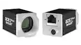
Two cameras for positioning and microscopic imaging
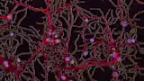
Tissue or blood samples on a sample holder are stained with fluorescent dyes to produce the microscope images. These dyes bind specifically to the cell or the tissue, which facilitates the examination of special areas in or on the cell. Depending on the staining technique used, the filtering of light, and the light emitted by the object, different problems can be worked on in the subsequent microscopic examinations.
SYNENTEC opted for two Basler ace GigE cameras. One of the cameras monitors the optimal position of the object in relation to the sample holder. The second camera is the main camera, delivering the actual microscopic image of the biological sample. A key feature is the camera sensor's high quality. When paired with the ability to select specific areas of the image (via AOI), a very high frame rate is generated.
With Basler, we have a partner who copes with complex tasks promptly and reliably, proving that customer service is far from an alien concept.
SYNENTEC is a leading manufacturer of high-throughput fluorescence and brightfield cell imaging systems and automation peripherals for cell based assays like in cell line development, upstream applications, drug discovery, cancer research, immunology, or stem cell research. SYNENTEC's objective is to provide superior devices for attractive and reasonable prices. The company is based in Elmshorn, Germany.