A Glimpse into the Living Cell: Nanolive's 3D Cell Explorer and Basler ace
- Customer
- NANOLIVE SA
- Location
- Ecublens, Switzerland
- Date
- 2015
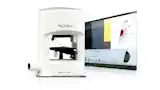
Nanolive SA has developed a microscope that, for the first time, makes it possible to study a living cell in 3D without damaging it. It offers unprecedented insights into the living cell. Using a Basler ace USB 3.0 camera, the 3D Cell Explorer immediately displays the cell in 3D, providing a comprehensive view of its morphology.
Actually see what happens inside a living cell
For the first time, the 3D Cell Explorer makes it possible to look inside a cell and observe internal features such as the nucleus and organelles. Thanks to the 3D Cell Explorer, scientists can actually see and precisely measure the effects of stimuli and drugs on cells.
High-precision light measurement in cells
Using a combination of holography and rotational scanning, the system detects changes in the light as it propagates through the cell. The sample is positioned between a high-numerical-aperture air objective below the sample and a rotating illumination arm above the sample. Green light (520 nm) from a diode laser is split into sample and reference beams. The sample beam illuminates the sample through the rotating illumination arm at a very steep angle.
Machine Vision: Precise Image Processing for Holographic Applications
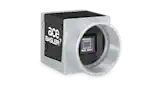
By combining the beam that has passed through the sample with the reference beam, the Basler ace USB 3.0 camera acA2000-165um then a hologram is recorded. The sample beam is then rotated by a small angle, and the process is repeated. A hologram is recorded for each beam position. This results in a complete image of the sample through the interference of the sample beam and the reference beam.
USB 3.0 CamerasSTEVE Software: Automatic and Intelligent Marking of Cell Regions
After a series of holograms has been captured, high-resolution images of each layer within the sample are generated through computer processing. Improved image resolution is achieved through the use of a synthetic aperture and multi-view holographic methods. The STEVE software developed by Nanolive makes it possible to highlight and label specific parts of the measured cells in 3D. The software automatically detects all regions with the same refractive index characteristics (different organelles have different optical properties) and digitally colors them with the same color.
The 3D Cell Explorer is a research tool, and we are just beginning to explore all its potential applications. It enables the measurement of cellular processes and kinetics in real time, thereby facilitating multi-parameter analysis at the cellular and subcellular levels.
About Nanolive
Nanolive SA is a startup founded in November 2013 at the EPFL Innovation Park in Lausanne, Switzerland. It has developed a revolutionary microscope that, for the first time, makes it possible to study a living cell in 3D without damaging it.