Microscopy Imaging Systems
Camera solutions for brightfield and fluorescence microscopy
Basler’s vision solutions enable precise, real-time visualization and analysis of microscopic structures by combining high-performance cameras with tailored software. Our systems deliver high-resolution imaging, accurate color reproduction, and high dynamic range, ensuring reproducible, detailed observation and analysis in applications such as histology, cell biology, hematology, and fluorescence imaging.
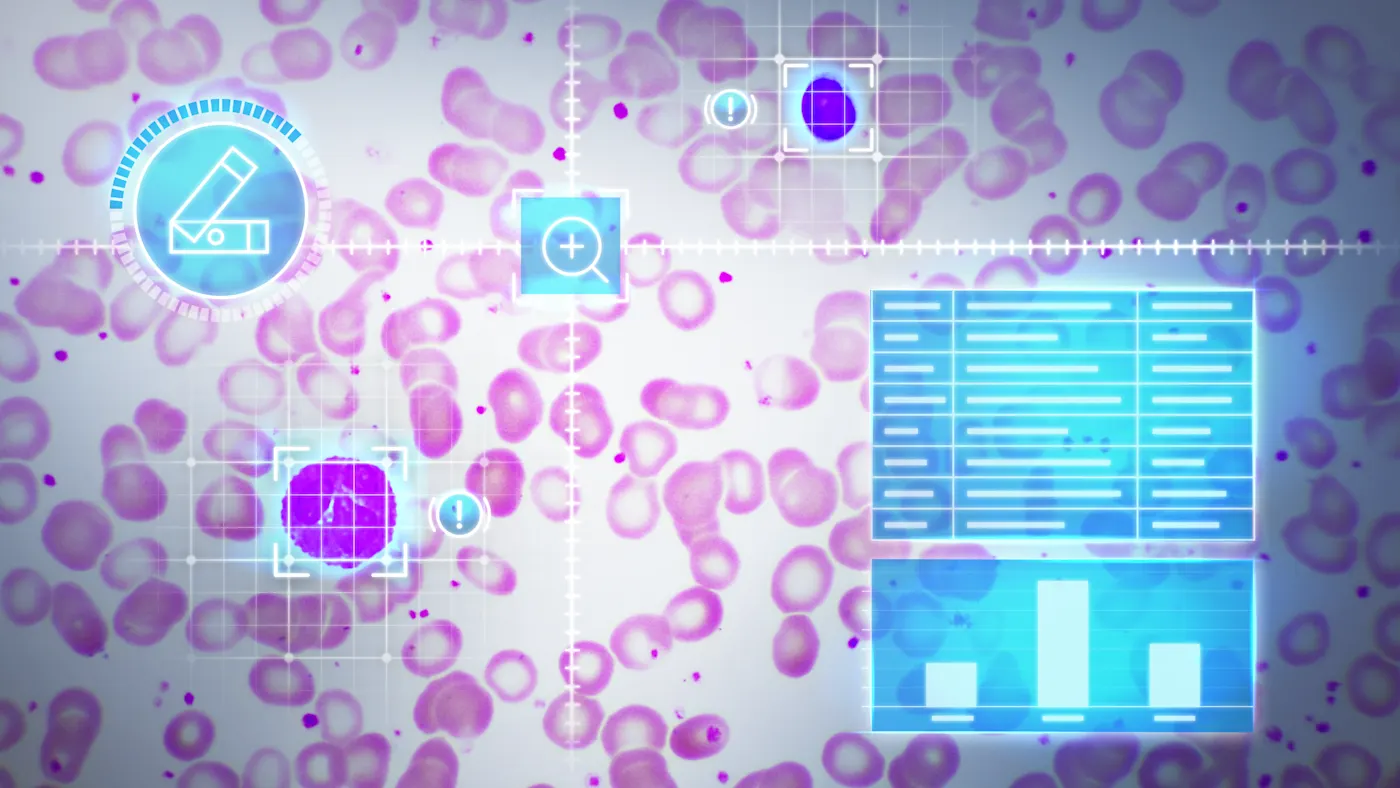
High image quality and color reproduction
Basler cameras enable high-resolution imaging and accurate color reproduction for detailed microscopy.Customizable hardware and software
Our project teams work with you to tailor vision components to your microscopy needs.Industry-leading reliability
Basler cameras have one of the lowest failure rates in the industry—built for reliable, repeatable processes in clinical diagnostics and research.Long-term product availability
Future-proof your system design with products that stay available and supported over time.
Key microscopy applications in life science research and clinical diagnostics
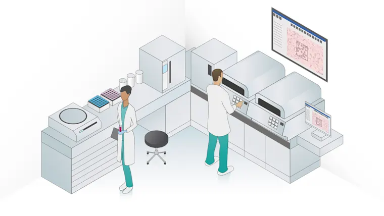
Vision solutions for image acquisition and processing are an integral part of microscopy. Light microscopy, including fluorescence microscopy, enables numerous applications in scientific, biomedical, and clinical laboratories.
Key applications include:
Histology
Digital pathology
Cell biology
Automated hematology
Medical microbiology
Conventional light microscopy
Many routine microscopic applications are based on light microscopy, using different illumination and contrasting techniques.
Basler cameras are a central component of these microscopy applications. They help track, record, analyze, and archive live images, delivering high-resolution and sharp images with accurate color fidelity.
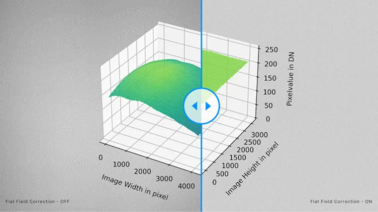
Light microscopy applications benefit from MED Feature Set "Brilliant Image"
“Brilliant Image" is one of the MED Feature Sets that is built into the Basler MED ace and is a customizable option for Basler ace 2 PRO. Learn about two features that help deliver the maximum image quality for your application.
Fluorescence microscopy
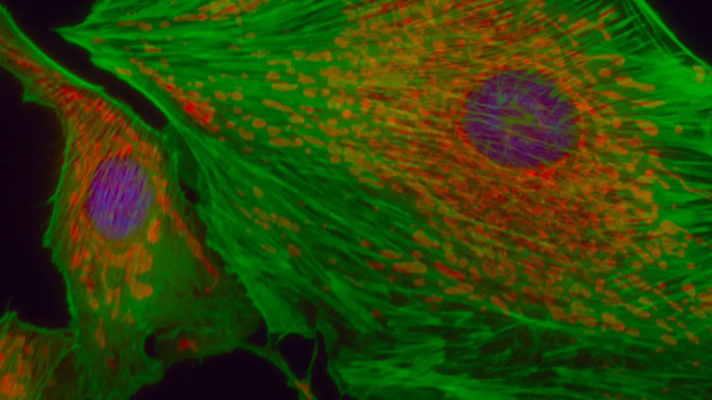
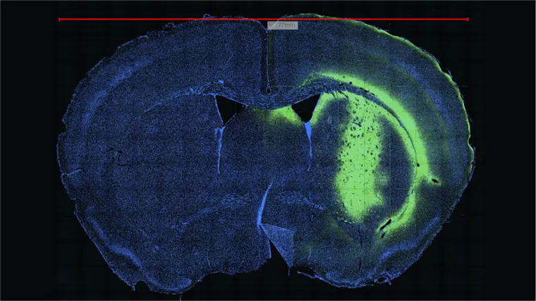
In fluorescence microscopy, a specific wavelength of light triggers fluorescent dyes to glow, allowing light emission detection. Even the most microscopic cellular and molecular structures can be detailed with fluorescence imaging.
Basler’s camera systems are critical for success. With minimal noise, a high dynamic range, and high light sensitivity, our cameras are ideally suited for fluorescence microscopy applications.
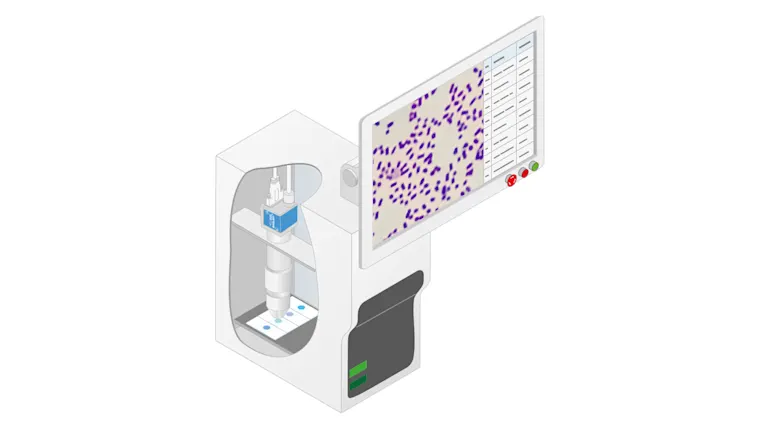
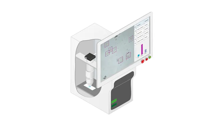
How Basler imaging solutions are used in fluorescence applications
Color calibration in Medical & Life Sciences
Digitally captured images contribute significantly to medical imaging systems and surgical devices, especially in ophthalmology, microscopy, and dermatology. In this context, color is often a decisive criterion for assessing whether a structure is healthy or diseased. Color fastness is therefore of utmost importance.

Why is color calibration important in medical technology?
In medical & life sciences, including ophthalmology, microscopy, or dermatology, the color fastness of digitally recorded images plays a critical role.
Read article
Do Basler cameras use color calibration?
Standardized color representation is important; it ensures that different imaging devices deliver the same result with the same color specifications. Learn color calibration step-by-step.
Download White PaperWhy should I color calibrate my camera?
For precise medical diagnoses, exact color reproduction in images and their color reproducibility are often indispensable. Color-calibrated cameras help with this.
How does the Basler Color Calibrator work?
Color reproduction is extremely important for images in Medical & Life Sciences. However, finding the perfect color settings can be complicated. Our Color Calibration Tool offers a simple solution.
Microscopy application use cases
There are several options that can be used to save time and money for a wide variety of medical and life science applications. Basler’s customized camera solutions can help.
Camera systems best suited to microscopy
Looking for detailed and efficient imaging in your microscopy system? The following Basler products are often the best choice:
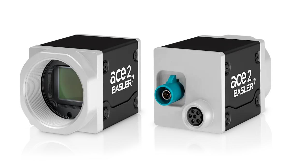
ace 2
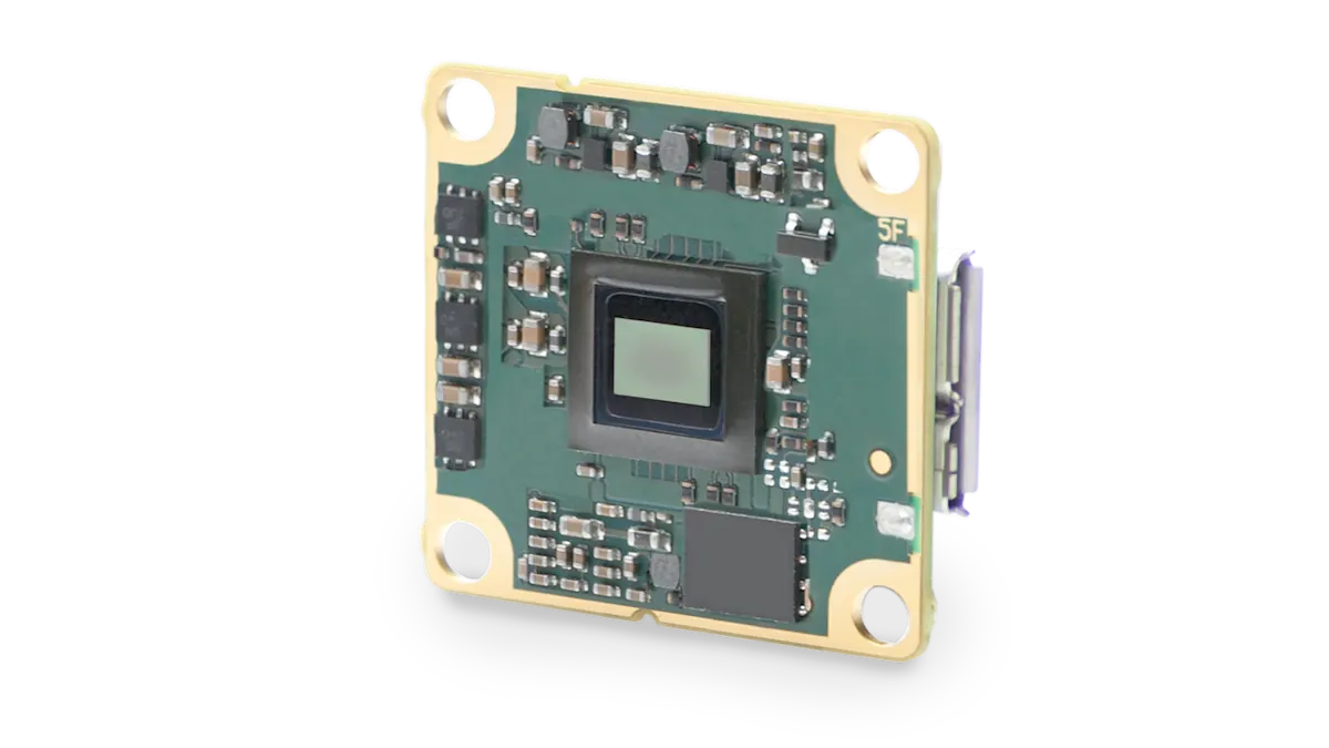
dart
racer 2
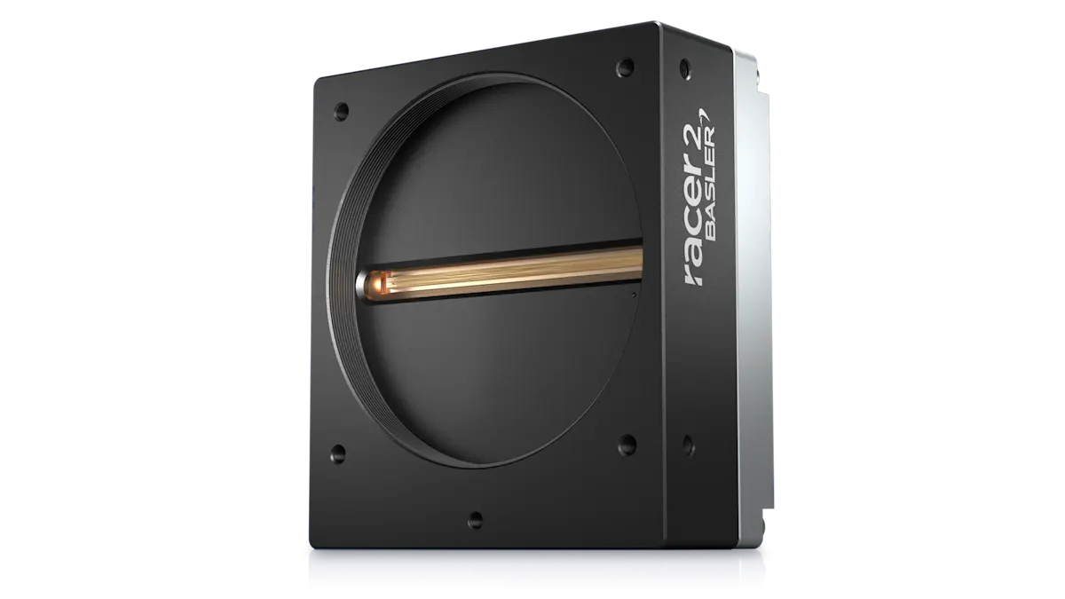
Area scan camera
Area scan camera
Line scan camera
Pixel resolution
Pixel resolution
Up to 25 MP
Pixel resolution
VGA to 13 MP
Pixel resolution
2k, 4k, 8k, 16k
Frame rate/line rate
Frame rate/line rate
Up to 190 fps
Frame rate/line rate
14 to 523 fps
Frame rate/line rate
Up to 240 kHZ
Interfaces
Interfaces
GigE, 5GigE, CoaXPress, USB 3.0, or GMSL2
Interfaces
GigE, USB 3.0, and BCON for MIPI
Interfaces
GigE, 5GigE, or CXP-12