AI Workflow for Automated Urinalysis
Automated sediment detection with CNN-based embedded vision systems
Urinalysis plays an important role in diagnosing kidney disease and urinary tract disorders. However, manual microscopy is slow, dependent on user skill, and often inconsistent. Laboratories require higher throughput, stable accuracy, and predictable costs without adding complexity to daily workflows.

Compact embedded vision for real-time sediment classification
Basler provides an embedded imaging solution that combines a Basler dart camera module, an NXP i.MX 8M Plus embedded board with integrated NPU and pylon AI software tools for automated urinalysis.
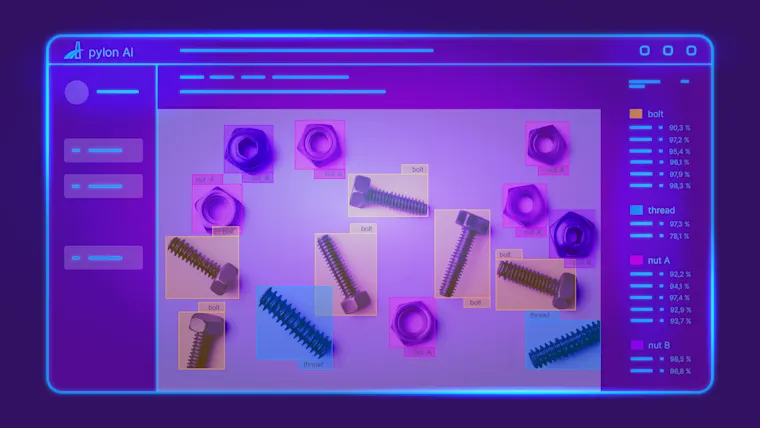
The system captures high-quality images of the sample, performs image preprocessing, and uses a trained convolutional neural network (CNN) to detect and classify relevant structures such as red and white blood cells, bacteria, casts, and crystals. The classification results are transferred to the host application for reporting and review.
How to use CNN-based imaging for urinalysis
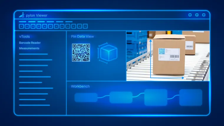
Image capture and preprocessing
The Basler dart camera acquires detailed images of the sample and performs preprocessing tasks such as delayering, denoising, and scaling to ensure consistent data quality.
Detection and classification with CNN
The trained CNN model runs directly on the NPU of the i.MX 8M Plus to identify red and white blood cells, bacteria, casts, and crystals in real time.
Visualization and output
The pylon Viewer displays bounding boxes and class labels, and the results are transferred to the laboratory information system (LIS) via standard interfaces for documentation and review.
Reliable AI results through consistent image data
Accurate sediment classification depends on stable and repeatable imaging results. The Basler dart camera, embedded processing, and pylon AI modules ensure consistent data quality and reliable AI inference.
This integrated approach enables laboratories and device manufacturers to achieve high throughput, consistent accuracy, and compact system integration while reducing total system cost through an embedded hardware-software platform.
Automate urinalysis with Basler’s camera systems
Manual microscopy shouldn’t limit throughput, consistency, or scalability. Basler’s CNN‑based embedded vision solutions enable reliable urinalysis and sediment classification, with repeatable performance and lower system complexity.
Build smarter lab automation workflows with BaslerProducts for this solution
Looking to implement a comparable solution? These products will help you.