Automated Microscopy for Blood Cell Analysis
High sample throughput rate with CNN-based imaging
Today’s microscopy produces massive volumes of image data, but extracting meaningful insights quickly and consistently remains a challenge. By combining automated microscopy with convolutional neural networks (CNN), laboratories can move beyond manual inspection and traditional rule-based image processing, enabling scalable, data-driven image analysis for complex biological samples.
What is high-throughput microscopy?
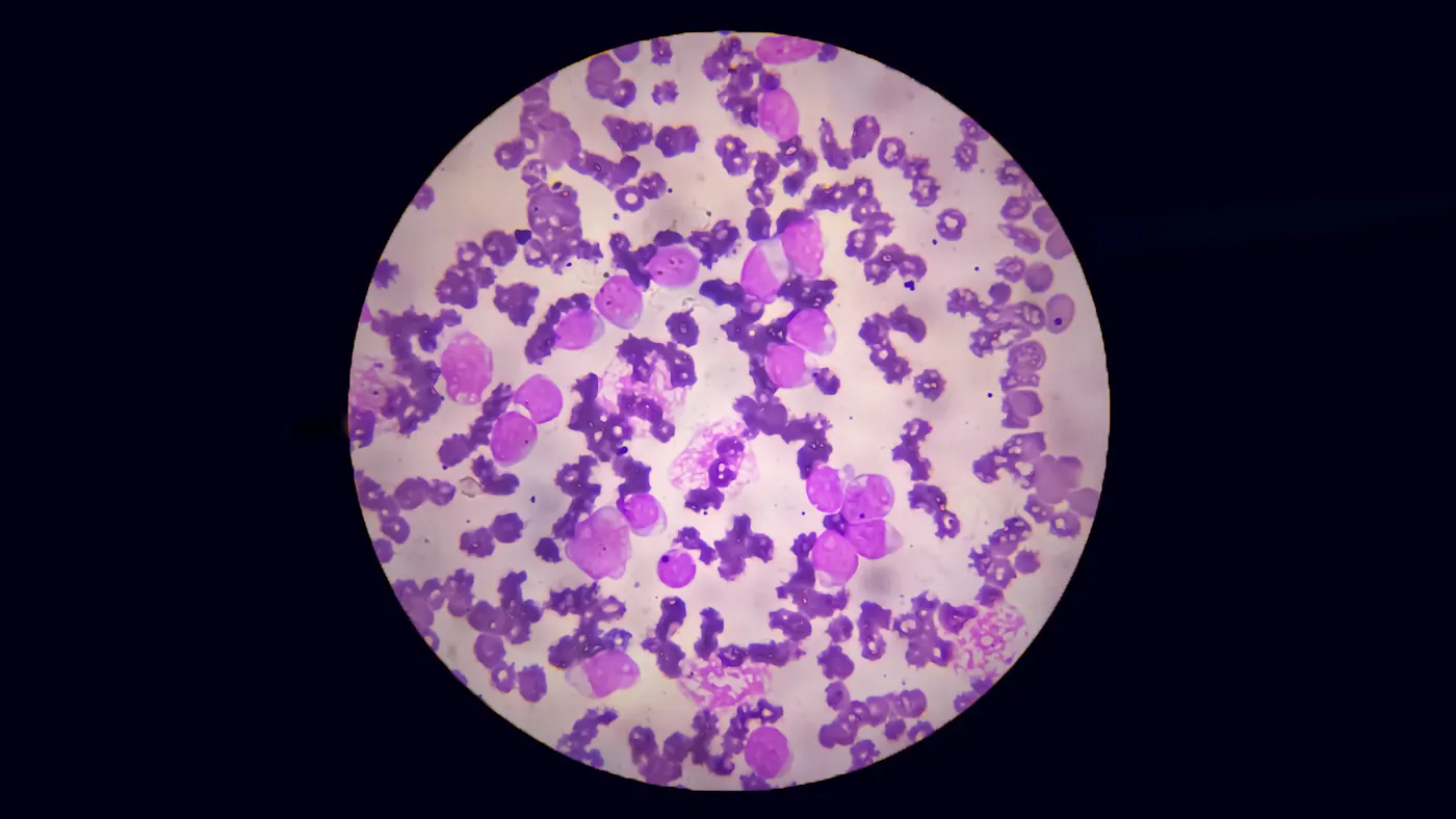
Which classes of blood cells are laboratory technicians seeing in a sample under a microscope? How many cells are there of each, and what are their sizes and shapes?
Answers to questions like these help pathologists diagnose diseases such as malaria, tuberculosis, or hematological-oncological disorders. However, using a manual microscope can be error-prone, time-consuming, and, ultimately, costly.
Enabling high-throughput screening while combating these factors, a CNN (Convolutional Neural Network) based computer vision can be used to automate this process.
What is the challenge in automated blood cell analysis?
Lab analyses of blood smears should be reliable, fast, and inexpensive. All of this is made possible by automating the analysis process.
The challenge starts here. Automation means high-resolution imaging and high-speed results. But it also means large amounts of data that must be transmitted and processed quickly.
Solving the automated microscopy problem with CNN-based computer vision
In our demo, we use a CNN to identify Plasmodium genera in blood smears, which can cause the infectious disease malaria. CNN sorts the Plasmodia into one of seven predefined classes and counts them. The result makes it possible to reliably determine the form of malaria.
To solve this or a similar problem, we will select the right hardware and software components for you and will configure them into a coherent computer vision system.
How to set up a CNN system for blood cell analysis
The system hardware in this demo includes a dual-channel Basler boost color camera with:
20 MP resolution
1.1” sensor format
CXP-12 interface
The camera enabled imaging at a high frame rate and resolution — a necessity for high scanning speed and sample throughput. But the (pre-)processing and analysis of large amounts of data required additional suitable components.
In this case, a programmable imaWorx CXP-12 Quad frame grabber handled not only the image processing and analysis but also supported autofocus and related features using FPGA-based real-time data pre-analysis. A C-mount lens and two CXP-12 data cables complete the system hardware, and Basler’s VisualApplets software was used to program the frame grabber FPGA.
The data pre-analysis programming and CNN implementation on the frame grabber FPGA are performed on the software’s graphic user interface. However, the frame grabber FPGA provides sufficient capacity for CNN implementation and enables high-performance inference.
Depending on the customer requirements, the CNN is either trained once and implemented on the frame grabber’s FPGA, or the customer is given the option to adapt the CNN later.
Advantages of Basler’s vision solutions for automated microscopy
High-quality imaging using the boost camera with Sony Pregius S Sensors
Optimized high-volume data processing with the 20 MP CXP-12 camera and frame grabber
Real-time data pre-analysis and CNN-based evaluation using over 900 MB/s
Reliable, high-throughput sample rate
Products for this solution
Looking to implement a comparable solution? These products will help you.