Lab Automation with Vision
Wide Range of Lab Applications
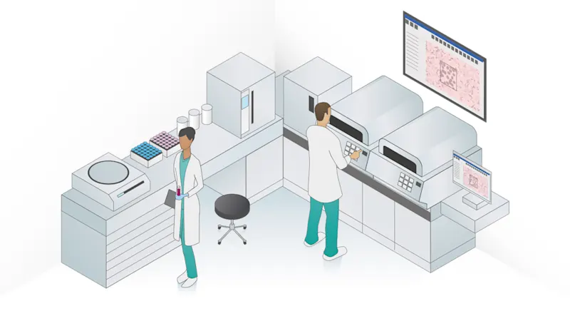
The concept of lab automation in general can be interpreted in many ways, and includes various tasks: from simple applications such as weighing, to complex robotic and analytical systems, process tracking, and storage systems. This results in numerous possible camera applications in the medical, scientific, pharmaceutical and analytical fields. Some of these are obviously recognizable, such as those in an imaging urine sediment analysis device, while others run in the background and provide information for the medical diagnosis we receive from a physician. Others in turn support processes that are internal to the devices, and not directly connected with the actual detection process. These applications range from the simple input of a barcode to the support of laser technologies; from the path traversed by blood, starting with the prick of the needle when the blood is drawn, to the various test processes and then the result, up to complex processes in cell technology which offer scientists insights into the origins of diseases, thus advancing diagnostic and therapeutic innovations.
Automation Trend in Medicine and Research
Hospital and research labs increasingly follow the trend towards automation. The essential drivers for this development are
1. Rising cost pressure:
Health systems and research institutions are subject to growing economic strains, and try to counteract this pressure with cost reductions in their services. Automation through modern technologies with inexpensive system components makes it possible to lower costs in the lab equipment, relieves the staff, and frees up capacities that can be utilized elsewhere.
2. High speed:
The faster processing of analyses enables more analyses per time spent for clinical and analytical contract laboratories, giving them an advantage over competitors since they can serve their customers faster. Automation can also help generate more results per time in research, which shortens project periods and makes new developments or technologies available sooner.
3. Better quality management and standardization:
Many examinations, once manually executed, are increasingly handled by machines, whose technological features make it possible to complete these tasks with greater precision and improved reproducibility. Thanks to an applied vision system and automated microscopy, for example, researchers can now view detailed and precise image data on their office monitors without having to look through eyepieces in darkrooms. Furthermore, the captured image data offers the capability of documentation and archiving, which meets the growing demands of quality management systems. Automated systems also aren’t subject to the process-related variances of manual work steps, giving them greater reproducibility and paving the way for advancing standardization. Digital image data can be viewed across different locations if desired, e.g. for a scientific exchange or an external diagnostic consultation. The conditions for a reliable diagnostic statement are therefore improved by camera-supported examinations and analyses.
4. Wider availability:
Lab automation efficiently makes new technologies accessible to many users. This makes it possible for research to determine the pathogenic processes of diseases more quickly. As a result,for example, diseases can be recognized earlier with the help of molecular-biological analyses in in-vitro diagnostics, which may reduce or even prevent their onset and the associated costly therapies that are so strenuous for patients. Devices that are easy to use and inexpensive enable diagnostics even in regions with economic- and infrastructure-related challenges. This means medical care can be improved in epidemic regions, since staff in those areas are often less well-trained, lab equipment has a lower standard overall, and the financial means of the affected patients are low. Here we can expect an increasing amount of so-called POC systems (POC = point of care) and lab-on-a-chip technologies.
Application Areas for Lab Automation
Below are some examples of typical application areas for automated, camera-based applications in labs:
1. Process automation
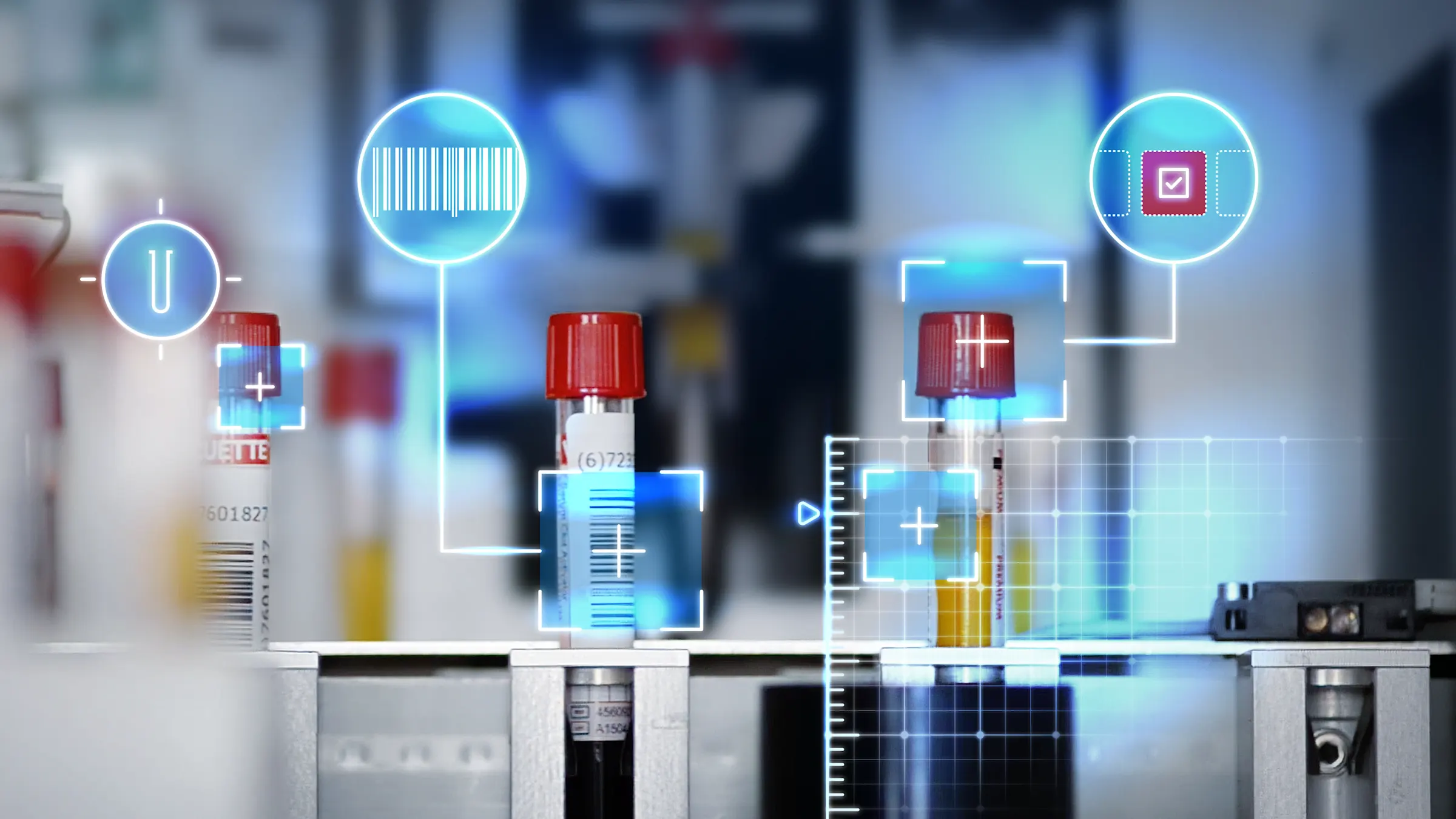
This includes general camera applications that generate imaging and data, not for purely analytical but for process-supporting purposes, e.g. barcode/matrix code compilation, as it is applied in most devices for in-vitro diagnostics (IVD). This could involve the simple identification of a patient’s sample vial or the transmission of data from the used reagent, which the device needs in order to calculate the analyses and a batch documentation for purposes of quality management. In an automatic exchange with a lab information system, the right results are thus attributed to the requirements of a patient sample and managed digitally.
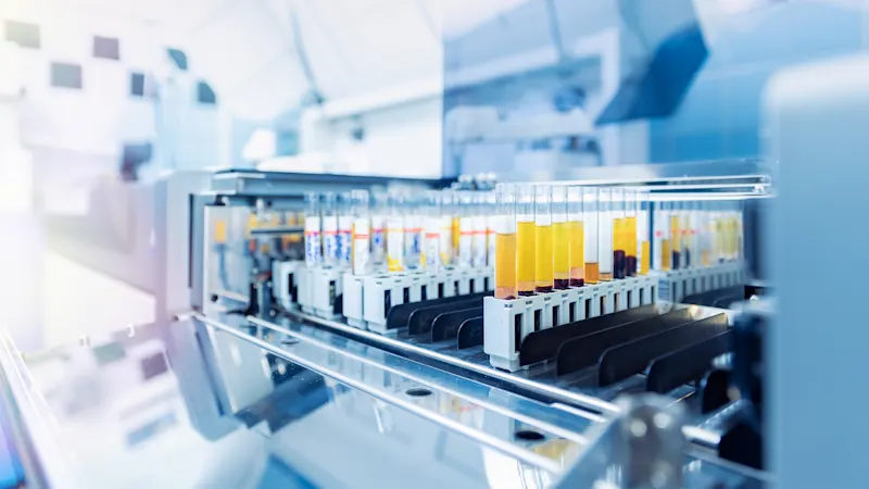
Many lab devices work with liquid test material. Depending on the application area, different parameters in this so-called liquid handling process must be determined and/or checked. This may be e.g. the state of the liquid (no air may be pipetted, since it would falsify the analysis result), the type of vial, the color of the lid to code the test material inside (e.g. whether it is a serum or whole blood vial), or color properties / layers or irregularities (bubbles, foam) in the liquid. Cameras may offer advantages since they don’t need contact with the sample, and don’t necessitate a removal of the lid, in contrast to other methods such as the capacitive determination of the liquid state. This prevents such problems as contamination, and enables higher flow rates.
2. Automated microscopy
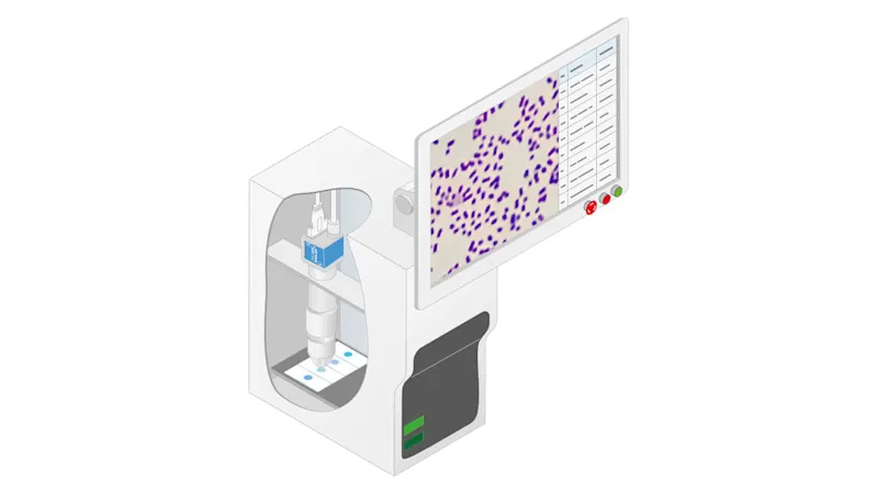
Automated microscopy includes, for instance, applications of light and fluorescence microscopy for in-vitro diagnostics (IVD), in life sciences, pharmaceutical research and in digital pathology.
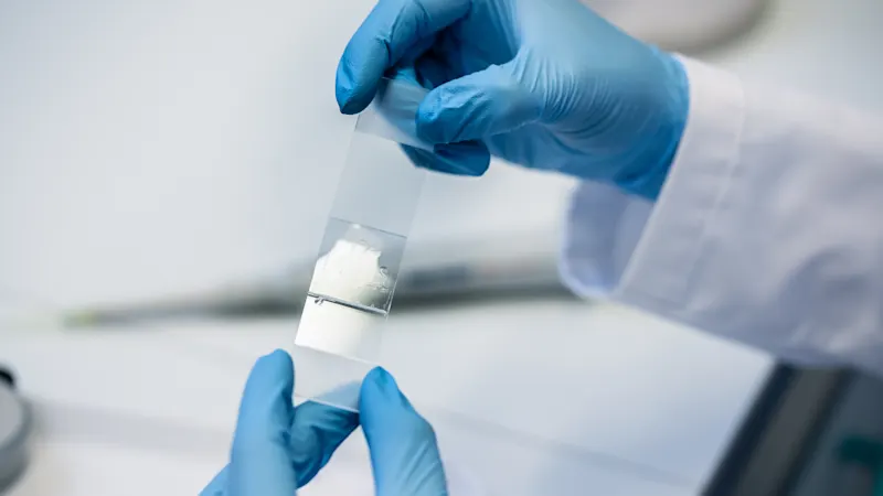
Different manufacturers use camera systems in their devices to diagnose autoimmune diseases, or diagnose diseases of the blood and hematopoietic organs in hematology, as well as in digital pathology. Pathologists examine tissue sections or cell samples for pathological changes. To this end, they prepare slides which can be examined by microscope to draw conclusions about diseases and provide valuable information for the diagnosis and therapy options, which may not be discernible through other means such as radiology.
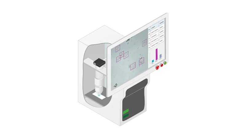
There is a wide selection of additional automatic microscope systems with different purposes. From a small device the size of half a shoe box, used for simple cell counting, to systems that are used directly in incubators and enable time-dependent life cell imaging without manual intervention, all the way to the high-content screening systems that are used e.g. in pharmaceutical substance screening – anything is possible.
Which Camera for Which Lab Application?
In addition to the above mentioned fields there is a wide variety of other potential applications and use cases of cameras in the whole scientific field, as for example in protein and nucleic acid analytics, microbiology, particle analytics and more. It’s important to offer cameras with the right features to cover a wide range of applications in the various specialty areas. Independently of the camera’s specific product features, it should offer easy and flexible integration with an efficient and comfortable SDK, and, of course, provide high quality and reliability. Technically excellent support, quickly and readily available, also simplifies the integration process for the system developer.
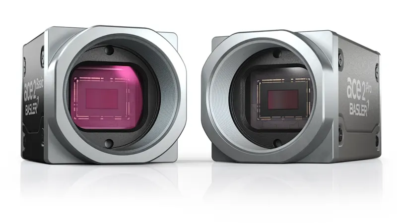
Nos produits pour le traitement d’images dans l’automatisation de laboratoire
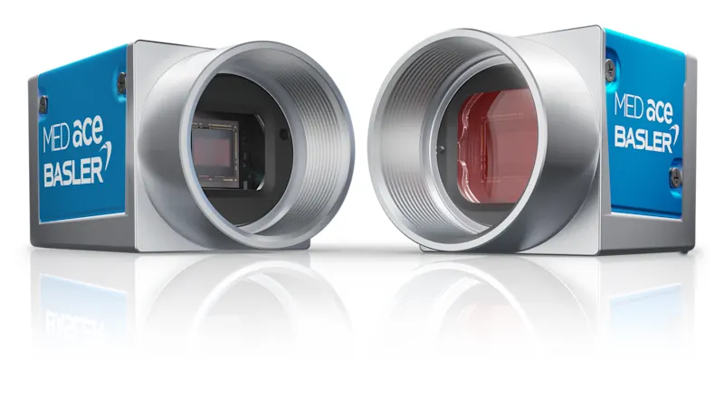
Profitez de la série spécialement conçue de caméras Basler MED ace et de notre expérience dans le développement d’une solution de vision industrielle individuelle adaptée à vos besoins.