Cameras for Fluorescence Microscopy OEM
Fluorescence microscopy is a very broadly used term covering numerous applications. This ranges from basic applications in the life sciences to highly developed techniques in which only very few photons or individual molecules are detected and localized by specific high-end hardware and software.
Last updated: 14/07/2023
Reading time: approx. 4 Minutes
CMOS sensors in fluorescence-based applications
In many systems, such as those used for medical, scientific, or clinical diagnostic applications involving fluorescence-based methods, CCD sensors need to be replaced by CMOS sensors. This white paper provides information on selecting a suitable camera and explains the related technical considerations.
To the fluorescence white paperWhy optical format and resolution matter
When you look at the visible wavelength range, the optical setup does not differ significantly from normal light microscopy applications in terms of format, magnification, and resolution. However, the overall cost can increase when larger optical formats, such as F-mounts, are used.
Key optical formats include:
Monochrome or Color
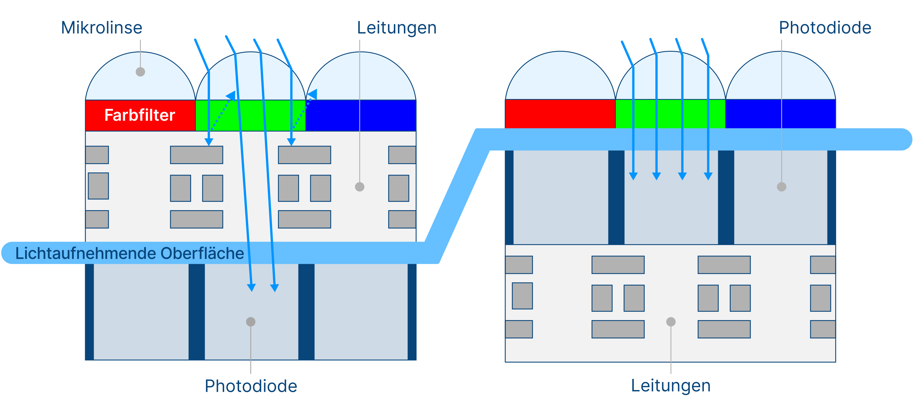
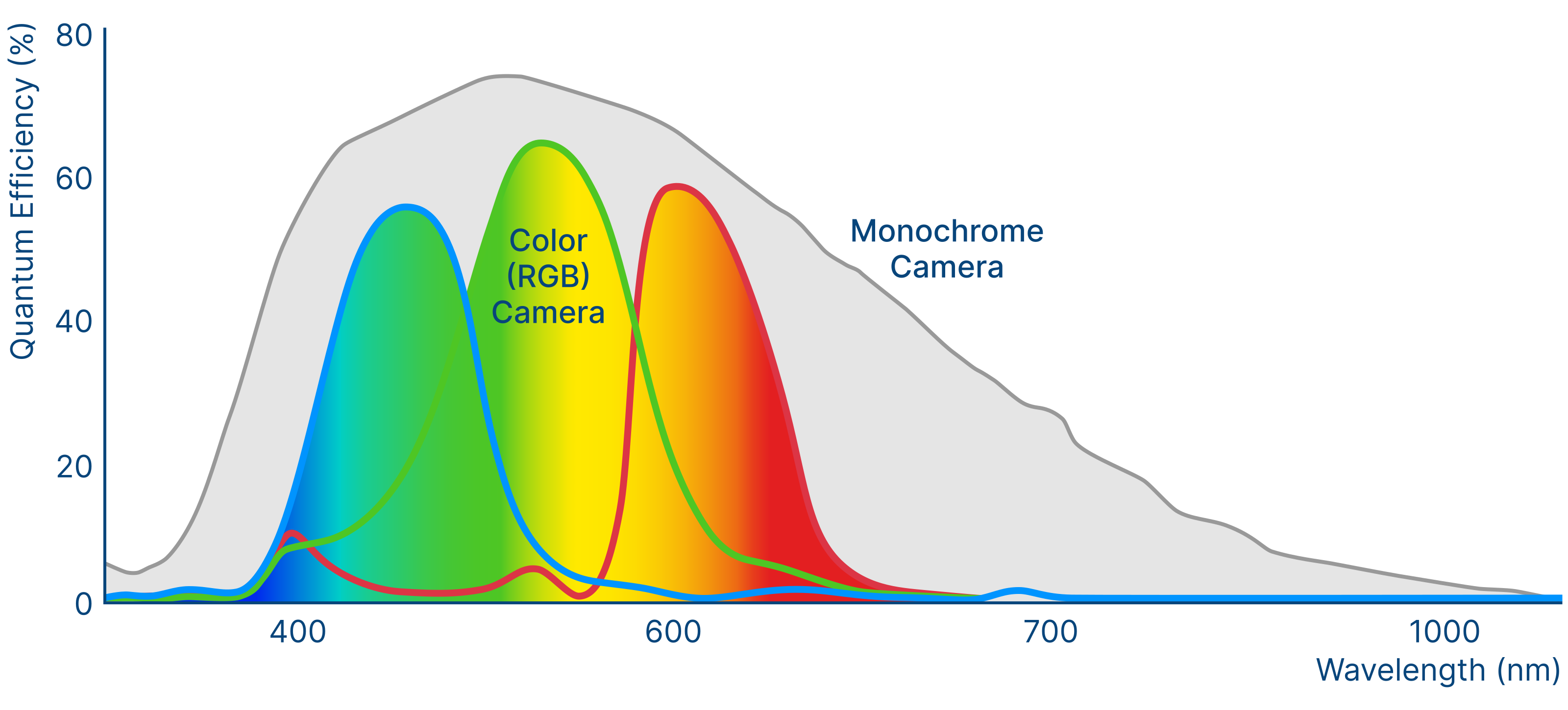
Monochrome cameras are generally preferred for fluorescence applications due to their higher quantum efficiency. The technical factor driving this difference is that in color cameras, Bayer microfilters on each pixel let only certain wavelengths pass through. This filtering is needed to calculate color information of the image using a process called debayering. As the color filters block a certain amount of light, fewer photons reach the photon-reactive area of the pixel. In addition to the Bayer pattern on the sensors, the IR-cut filter in color cameras presents a limiting factor because it blocks light of approximately 650 to 700 nm upward.
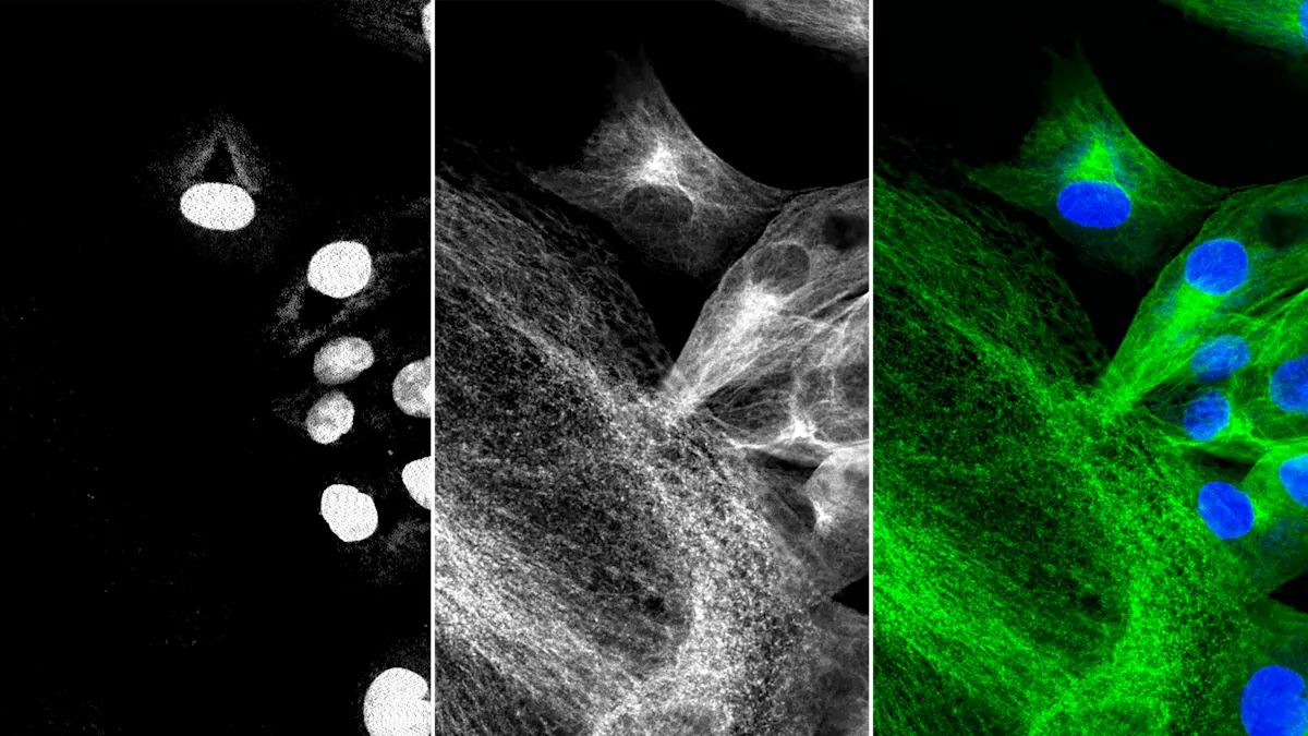
Typically, images with multiple fluorescence markers for specific detection and co-localization of molecules of interest are made from separate images using monochrome cameras. Selectable light sources and filter sets provide the right combination of excitation and emission wavelengths for each fluorophore used.
However, certain applications may create a demand to do color imaging and fluorescence within one instrument using only one camera. This is possible if the sensitivity demands of the fluorescence application are not too high.
Fluorescence in the Field
Fluorescence is a physical phenomenon and not just a specific technology. The possible methods – e.g. for analytics, quantitative determinations or visualizations used in the life sciences – are almost infinite. Fluorophores can be coupled to various carriers such as proteins (often antibodies), nucleic acids or microparticles. But they can also be integrated as gene technology markers in organisms in order to examine cell-biological functions and processes. Aside from the life sciences, fluorescence-based methods are also used in other areas, such as material analysis or forensics. The following examples show the versatile application options for fluorescence.